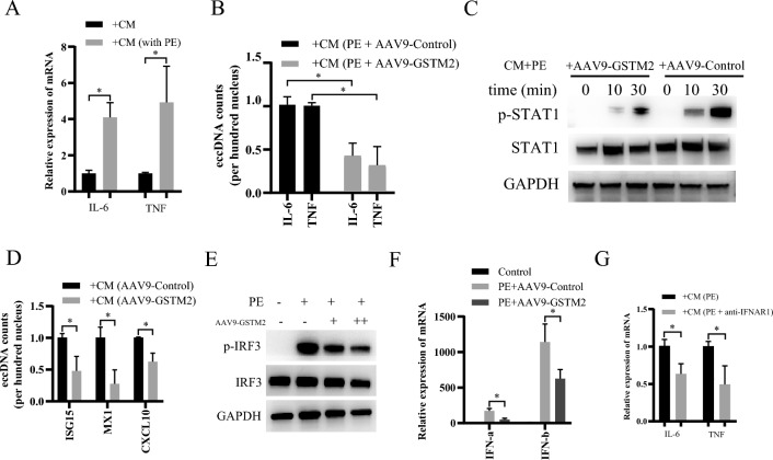
Fig. 7.
GSTM2 alleviates IFN-I-stimulated macrophage inflammation. A qRT‒PCR analysis of IL-6 and TNF-α mRNA expression in RAW264.7 macrophages treated with culture supernatants from PE- or DMSO-treated cardiomyocytes. n = 3. *p < 0.05. B qRT‒PCR analysis of IL-6 and TNF-α mRNA expression in the RAW264.7 macrophages treated with culture supernatants from PE-treated cardiomyocytes infected with AAV9-GSTM2 or control AAV9. n = 3. *p < 0.05. C Western blotting analysis of STAT1 and p-STAT1 expression in RAW264.7 macrophages treated with culture supernatants from PE-treated cardiomyocytes infected with AAV9-GSTM2 or control AAV9. GAPDH was used as a loading control. D qRT‒PCR analysis of ISG15, MX1, and CXCL10 mRNA expression in RAW264.7 macrophages treated with culture supernatants from PE-treated cardiomyocytes infected with AAV9-GSTM2 or control AAV9. n = 3. *p < 0.05. E Western blotting analysis of IRF3 and p-IRF3 expression in the cardiomyocytes treated with PE alone or combined with AAV9-GSTM2 infection. GAPDH was used as a loading control. F Assessment of IFN-α and IFN-β levels in the culture supernatants from PE-treated cardiomyocytes infected with AAV9-GSTM2 or control AAV9. n = 3. *p < 0.05. G qRT-PCR analysis of IL-6 and TNF-α mRNA expression in RAW264.7 macrophages treated with culture supernatants from PE- and IFNAR1 antibody-treated cardiomyocytes. n = 3. *p < 0.05

