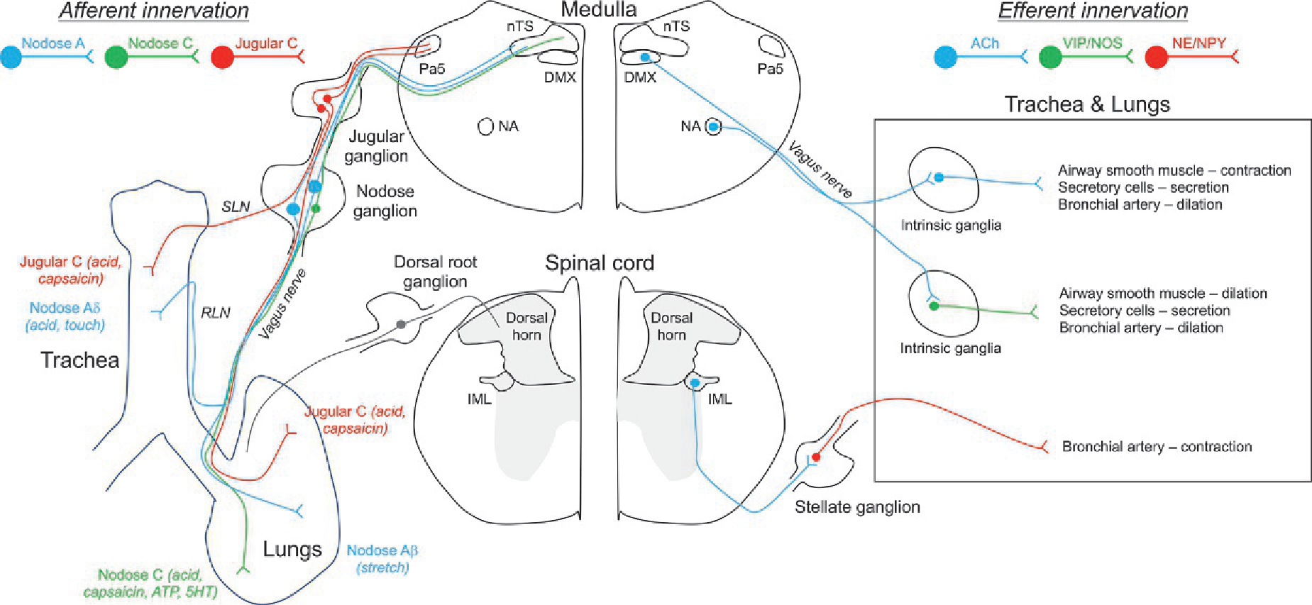
Fig. 14.1.

Schematic illustrating the basic sensory afferent and autonomic efferent innervation of the airways. Left, sensory fibers projecting from neurons within the nodose and jugular ganglia project to the trachea and intrapulmonary airways, via the vagus nerve and its branches the superior laryngeal nerve (SLN) and the recurrent laryngeal nerve (RLN). Sensory afferents have been grouped based upon their conduction velocity and their sensitivity to specific stimuli. Nodose sensory fibers project to the nTS in the medulla and jugular fibers project to the Pa5 in the medulla, where they synpase with second order neurons involved in networks regulating cardiopulmonary reflexes. Also shown is the sparse sensory innervation projected from the dorsal root ganglion. Little is known about the sensitivity profile, anatomical pathways and function of these DRG afferents, but it is likely that many are C fibers that project to the dorsal horn lamina. Right, extrinsic and intrinsic autonomic innervation of the airways. Cholinergic preganglionic parasympathetic fibers project from the dorsal motor nucleus of the vagus (DMX) and nucleus ambiguus (NA) to the airways where they synapse with neurons within the intrinsic ganglia. Such postganglionic fibers are either cholinergic or NANC fibers (expressing VIP and/or NOS), and they project to airway smooth muscle cells, secretory cells and bronchial arteries. Cholinergic preganglionic sympathetic fibers project from the intermediolateral nucleus of the spinal cord to neurons within the stellate and thoracic sympathetic ganglia. These adrenergic and NPY-expressing ganglionic neurons project to the airways and mainly innervate bronchial arteries.
