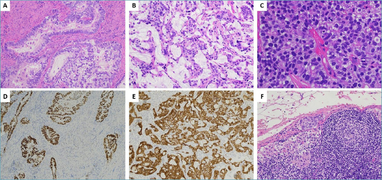
Figure 2.

Histological and immunohistochemical findings observed in the surgical specimen. (A) Neoplastic glands were composed of mucinous cells with voluminous clear cytoplasm and distinct cell borders. (B) Neoplastic glands were haphazardly distributed, showed moderate nuclear atypia and variable amounts of intracytoplasmic eosinophilic mucin. (C) Higher magnification (x 20), demonstrating a solid pattern of growth with neoplastic cells showing pale eosinophilic or clear cytoplasm and moderate nuclear atypia. (D) Immunohistochemistry confirmed the diffuse for CDX2 also in the surgical sample. (E) Diffuse staining for p16 was also observed. (F) Microphotograph illustrating isolated tumor cells (ITCs) detected in sentinel lymph-node.
