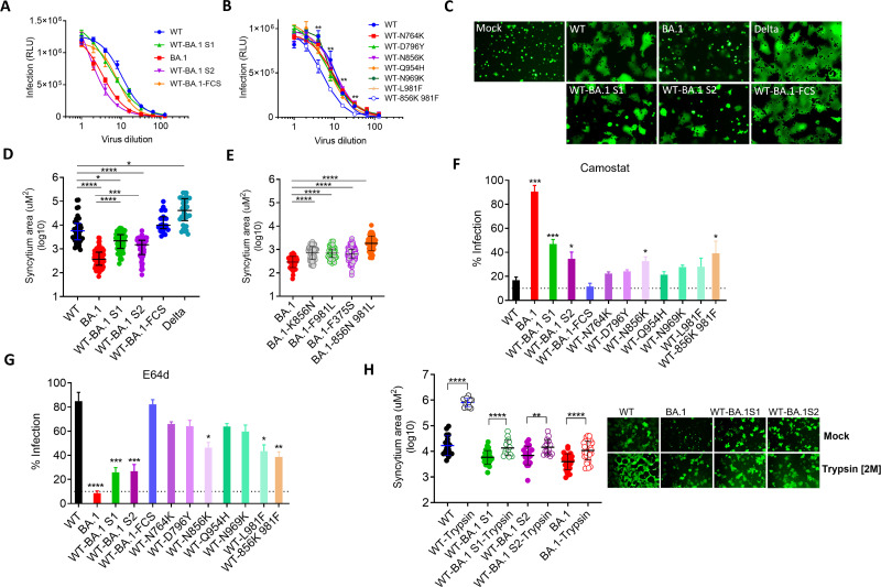
Fig 7.
Entry of PVs bearing various spike proteins: (A and B) Serially diluted PVs were used for the infection of 293T-hACE2-TMPRSS2 cells. Infectivity [relative luminescence unit (RLU)] is shown against various dilutions of PV expressing parental and mutant spikes. The statistical significance for the difference in infectious titers between WT and WT-856K 981F at a given dilution was calculated using Mann-Whitney test. (C) HEK293T cells expressing Spike and GFP were added onto the monolayer of Vero-TMPRSS2. Shown are the images of syncytia after 2-hour incubation formed by Spike variants, or without Spike (Mock). (D and E) The size of the syncytia measured in µm2 (micrometer squared) plotted for indicated Spike variants. (F and G) Infectivity of PVs in the presence of TMPRSS2-inhibitor camostat or cathepsin-inhibitor E64D. The statistical significance was calculated with reference to WT. The statistical significance in D–G was calculated using Kruskal-Wallis test, and comparisons were done by Dunn’s multiple comparison post test. (H) Syncytia formation of Vero-TMPRSS2 by Spike-expressing HEK293T cells in the presence of trypsin. The images were taken after 2 hours of addition of spike-expressing 293T cells onto Vero-TMPRSS2. The statistical analysis was done by using Kruskal-Wallis test, and P values were calculated by Dunn’s multiple comparison post-test. All the experiments were repeated at least twice.

