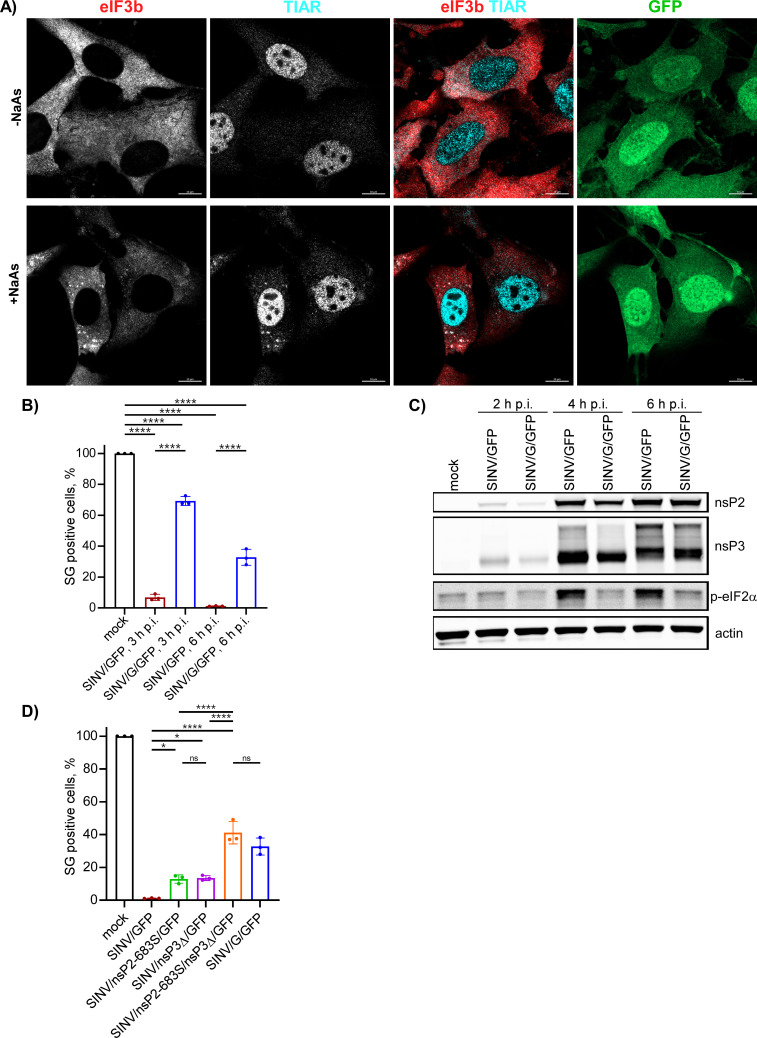
Fig 13.
The inabilities of viral mutants to develop transcriptional or translational shutoffs make them incapable of blocking SG development in response to NaAs treatment despite the accumulation of wt nsP3. (A) NIH 3T3 cells were infected with SINV/G/GFP at an MOI of 10 PFU/cell. At 6 h p.i., they were treated with 0.75 mM NaAs for 45 min or remained mock treated. Cells were fixed, immunostained with antibodies specific to the indicated SG markers, and analyzed by confocal microscopy. Bars correspond to 10 µM. (B) NIH 3T3 cells were infected with the indicated viral variants at an MOI of 10 PFU/cell. At the indicated times p.i., cells were treated with 0.75 mM NaAs for 45 min and immunostained with TIAR- and eIF3b-specific antibodies to detect SGs. The percent of GFP-positive cells containing SGs was determined (~100 cells per experiment). Means and SD are indicated. The significance of differences was determined by one-way ANOVA with the two-stage step-up method of Benjamini, Krieger, and Yekutieli test (****P ≤ 0.0001, *P ≤ 0.05, ns ≥0.05; n = 3). (C) NIH 3T3 cells in six-well Costar plates were infected with SINV/G/GFP and parental SINV/GFP at an MOI of 10 PFU/cell and harvested at the indicated times p.i. Cell lysates were analyzed by WB for accumulation of viral nsPs and p-eIF2α. (D) NIH 3T3 cells were infected with the indicated viral variants at an MOI of 10 PFU/cell. At 6 h p.i., they were treated with 0.75 mM NaAs for 45 min with TIAR- and eIF3b-specific antibodies. The percent of GFP-positive cells containing SGs and statistical analysis was performed as described above for panel (B).

