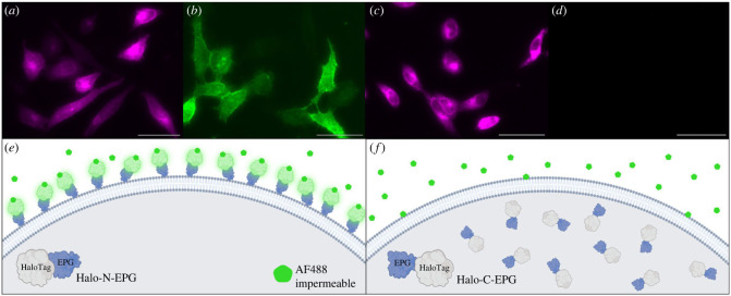
Figure 1.
Fluorescently labelled HaloTag indicates EPG's cellular localization. (a–d) HeLa cells expressing Halo-N-EPG (a,b) and Halo-C-EPG (c,d) labelled with fluorescent HaloTag ligands: (a,c) membrane permeable JFX650, and (b,d) membrane impermeable AF488. (e,f) Illustrations to demonstrate the interaction of HaloTag-EPG fusion proteins with AF488. (e) As reflected in (b), Halo-N-EPG expressing cells were labelled with AF488 indicating its presence as a membrane associated protein with its N terminus exposed to the extracellular space. (f) As reflected in (d), Halo-C-EPG expressing cells were not labelled with AF488, indicating this construct does not localize to the membrane. Scale bars represent 50 µm.

