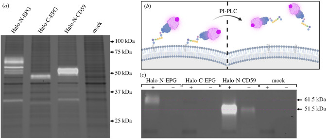
Figure 2.
Lysate analysis and PI-PLC digestion signify EPG undergoes posttranslational modification to receive a glycosylphosphatidylinositol anchor. (a) Lysate of HeLa cells expressing HaloTag fusion proteins labelled with JFX650 run on an SDS-PAGE gel visualized with Far-Red excitation and 715/30 filter emission (white bands), and Stain-Free imaging (black bands). The expected size of the HaloTag fusion proteins is approximately 46 kDa. Halo-N-EPG presents as a series of bands between 50 and 75 kDa. Halo-C-EPG exhibits a band at 46 kDa. Halo-N-CD59 presents a band just above 50 kDa. Mock transfected cells do not present JFX650 associated bands. (b,c) HeLa cells expressing HaloTag fusion proteins labelled with JFX650 were treated with PI-PLC, effectively releasing any GPI anchored protein from the membrane as illustrated by (b). Media from treated (+) and untreated (−) cells visualized by SDS-PAGE with Far-Red excitation and 715/30 filter emission (white bands) and Stain-Free imaging (black bands) show a band at approximately 61.5 kDa in the Halo-N-EPG (+) group that is not present in the untreated (−) group—indicative of the presence of a GPI anchor. Halo-C-EPG did not present any JFX650 associated bands. The positive control, Halo-N-CD59, presented bands at approximately 51.5 kDa. Mock transfected cells did not present any JFX650 associated bands.

