Figure 3.
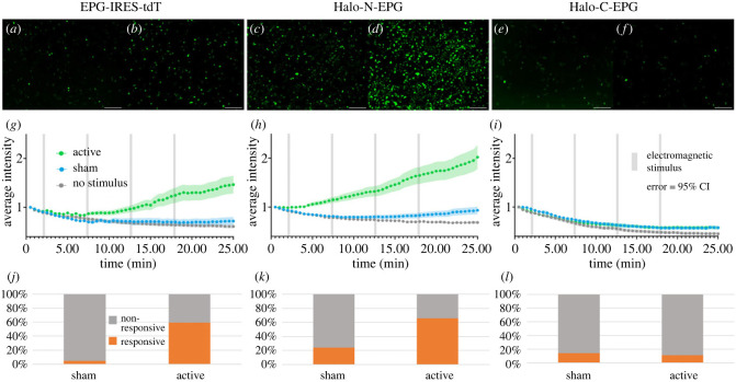
Developing an assay using GCaMP6m to determine if EPG is still functional after the addition of HaloTag. (a–f) HeLa cells expressing GCaMP6m and various EPG-HaloTag fusion constructs visualized before (a,c,e) and after (b,d,f) electromagnetic stimulation. (a,b) Cells expressing EPG-IRES-tdT appear more intense after stimulation. (c,d) Cells expressing Halo-N-EPG appear more intense after stimulation. (e,f) Cells expressing Halo-C-EPG appear relatively unchanged after stimulation. Scale bars represent 200 μm. (g–i) Average intensity of GCaMP6m over time with various electromagnetic stimuli. Error bars are representative of 95% CI. Significant increases in intensity were observed between the no stimulus/sham and active groups in both (g) (p < 0.0001, unpaired t test) and (h) (p < 0.0001, unpaired t test). No significant difference was observed between any groups in (i). (j–l) Percentage of individual cells that produced a signal greater than of the corresponding no stimulus group. Significant differences were observed between sham and active groups in both (j) and (k). No significant difference was observed between sham and active groups in (l). The EPG-IRES-tdT group included n = 177, n = 134, and n = 163 cells over four experiments for no stimulus, sham, and active groups respectively. The Halo-N-EPG group included n = 282, n = 269, and n = 279 cells over four experiments for no stimulus, sham, and active groups respectively. The Halo-C-EPG group included n = 160, n = 182, and n = 189 cells over three experiments for the no stimulus, sham, and active groups respectively.

