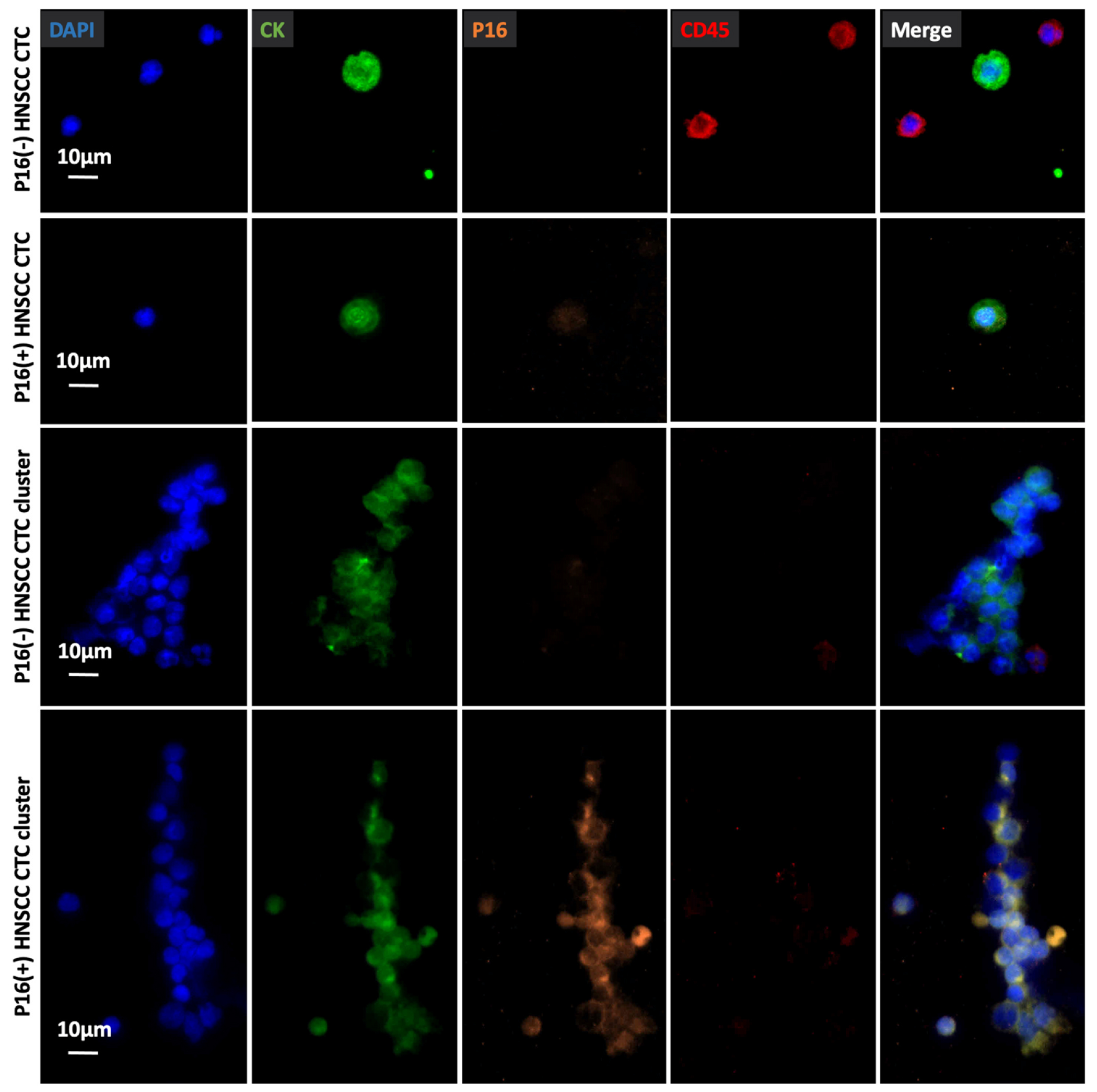
Fig. 6.

Representative micrographs of an immunocytochemistry (ICC)-stained P16(−) HNSCC CTC, P16(+) HNSCC CTC, 16(−) HNSCC CTC cluster, and a P16(+) HNSCC CTC cluster on TDN-NanoGold Click Chips from different HNSCC patients. Blue: DAPI stained nuclei; green: FITC stained CK; orange: TRITC stained P16; red: CY5 stained CD45. Scale bar, 10 μm. Data are representatives of five independent assays.
