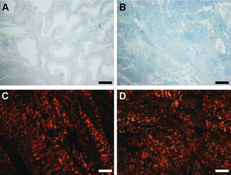
Fig 3. Histology ECM components.
A) Decellularized healthy tissue, stained with Masson Trichrome (MT). B) Decellularized fibrotic tissue, stained with MT. C) decellularized healthy tissue, stained with Picrosirius Red (PR). D) Decellularized fibrotic tissue, stained with PR. A-B scale bar represents 200 μm, panels C-D scale bar represents 40 μm.

