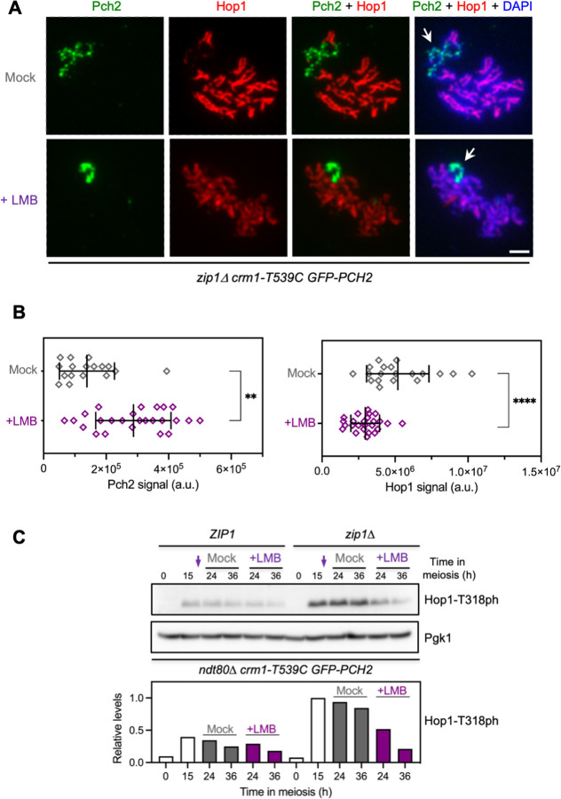
Fig 2. LMB treatment impairs the meiotic recombination checkpoint.
(A) Immunofluorescence of zip1Δ spread meiotic chromosomes stained with anti-Hop1 (red) and anti-GFP (to detect GFP-Pch2; green) antibodies. Representative nuclei, either mock-treated, or treated with 500 ng/ml LMB 15 h after meiotic induction, are shown. Arrows point to the rDNA region. Spreads were prepared at 19 h. Scale bar, 2 μm. The strain is DP1721 (zip1Δ crm1-T539C GFP-PCH2). (B) Quantification of Pch2 (left graph) and Hop1 (right graph) signal for the experiment shown in (A). Error bars, SD; a.u., arbitrary units. (C) Western blot analysis of Hop1-T318 phosphorylation (ph) in the absence (mock) or presence of LMB added at 15 h after meiotic induction (arrows). Pgk1 was used as loading control. The graph shows the quantification of Hop1-T318ph relative levels. Strains are: DP1927 (ndt80Δ crm1-T539C GFP-PCH2) and DP1837 (zip1Δ ndt80Δ crm1-T539C GFP-PCH2).

