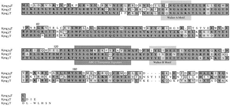
FIG. 5.
CLUSTAL alignment of the predicted amino acid sequences of E. coli kpsK54T (this study), kpsK5T, and kpsK1T. The boxed sequence identifies amino acid residues that are functionally similar (lighter shading) or identical (darker shading). The ATP-binding domain Walker A (residues 38 to 45), and Walker B (residues 145 to 151) motifs (34) and the ABC-2 transporter signature sequence (residues 125 to 139) are marked (1, 18). Numbers above the sequences are residue numbers.

