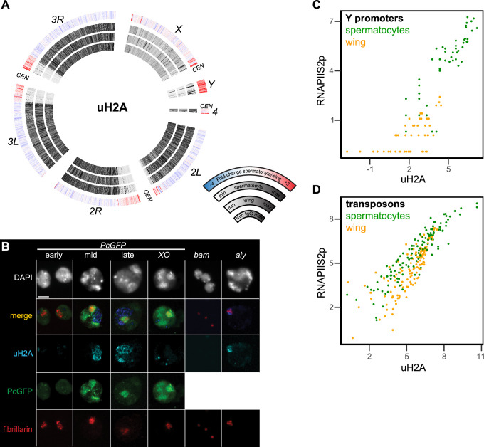
Figure 6. Chromosomal distribution of ubiquitinylated histone H2A in isolated spermatocytes.
(A) CIRCOS plots of uH2A across Drosophila chromosomes in IgG controls, in wing imaginal discs, and in isolated spermatocytes. (B) Immunostaining of uHA (blue) and the nucleolar marker fibrillarin (red) on germline nuclei. Early-, mid-, and late-spermatocyte stages were identified by Polycomb-GFP (PcGFP) (green) localization pattern in wildtype spermatocytes and in X/O spermatocytes. Testes from bam mutants contain gonial cells, while testes from aly mutants contain mostly early spermatocytes. All images are displayed at the same magnification, and a 100 µm scale bar is shown in the top left section. (C) Correspondence of uH2A and RNAPIIS2p signals around the promoters of genes on the Y chromosome in wing imaginal discs and in isolated spermatocytes. (D) Correspondence of uH2A and RNAPIIS2p signals across transposon consensus sequences in wing imaginal discs and in isolated spermatocytes.


