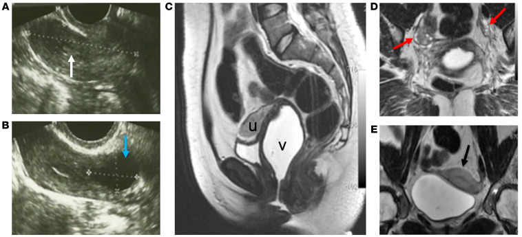
Figure 1. FRT imaging features of a 29-year-old woman (proband) with primary amenorrhea and a developmental anomaly of the reproductive tract.
(A and B) Transvaginal ultrasonographic imaging showing (A) a hypoplastic uterus (dotted line) with a very thin endometrium (white arrow), and (B) a left cornual cystic lesion (blue arrow); (C–E) Sagittal (C), oblique coronal (D), and coronal (E) T2-weighted fast-recovery fast spin-echo (FRFSE) MRI showing pelvic sections with vaginal distention using aqueous gel, demonstrating: (C) widening of the upper 2 thirds of the vagina (v) and an anteverted hypoplastic uterus (u), (D) normal appearing ovaries (red arrows), and (E) a hypoplastic uterus with thin endometrium (black arrow). The MRI of the proband was performed while off of hormonal treatment for more than 1 year. No information about cycle phase was available since the patient was amenorrheic.

