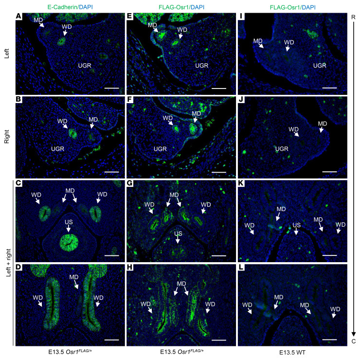
Figure 4. Osr1 expression in the MDs of E13.5 Osr1FLAG/+ mouse embryos.
(A–D) Cross-sections of urogenital ridges showing WDs, distinguished from MDs by staining for E-cadherin (anti-E-cadherin antibody, green), which is an epithelial marker specifically expressed in WDs, but not in MDs, at E13.5. (E–H) Osr1 is expressed (anti-FLAG antibody, green), in MDs, as well as in WDs, throughout their entire length—i.e., from rostral to caudal segments. (I–L) Sections of WT E13.5 embryos at the same levels were used as negative controls (anti-FLAG antibody). UGR, urogenital ridge; US, urogenital sinus; R, rostral; C, caudal. Scale bars: 100μm.

