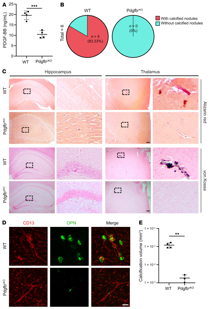
Figure 3. Deletion of Pdgfb from bone preosteoclasts alleviates brain calcification.
(A) ELISA measurement of serum PDGF-BB concentrations in 17-month-old PdgfbcKO male mice and WT littermates. n = 4–5. (B) Calculation of the calcification incidence in PdgfbcKO mice and WT littermates based on histology. n = 6. (C) Representative images of Alizarin red– and von Kossa–stained images for 17-month-old PdgfbcKO mice and WT littermates. Calcification nodules are shown in red and black, respectively. Boxed areas are shown at higher magnification (×40) in the corresponding panels on the right. n = 6. (D) Double-immunofluorescence staining of frozen brain tissue sections from 17-month-old PdgfbcKO mice and WT littermates using antibodies against CD13 and OPN. n = 3–4. (E) Quantification of the volume of OPN+ calcified nodules in D. n = 3–4. The Imaris 3D reconstruction method was used to quantify the number and volume of calcification. Scale bars: 100 μm (C and D). Data are shown as the mean ± SD. **P < 0.01 and ***P < 0.001, by unpaired, 2-tailed Student’s t test (A and E).

