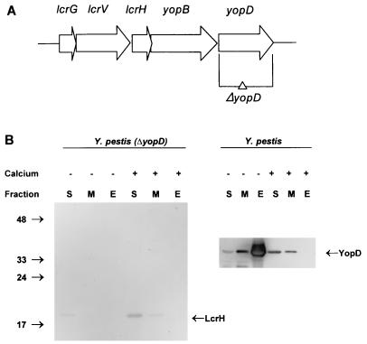
FIG. 1.
Construction of a complete ΔyopD mutation in Y. pestis. (A) Schematic representation of the lcrGVHyopBD operon of Y. pestis LCR plasmid pCD1. The positions and directions of the genes of this operon are shown as horizontal arrows. The DNA region deleted to create the yopD mutation is indicated (ΔyopD). (B) Immunoblot analysis of YopD and LcrH expressed in Y. pestis KIM8-3002.2 (ΔyopD) and Y. pestis KIM8-3002 (parent). Bacteria were grown at 37°C in the absence (−) or presence (+) of 2.5 mM Ca2+. Proteins from equal numbers of cells (0.025 A620 unit · ml) were separated by SDS-PAGE in a 15% (wt/vol) polyacrylamide gel. Antibodies were used to detect YopD and LcrH in the soluble (S) (cytoplasmic and periplasmic) fraction, the membrane (M) fraction, and the extracellular (E) fraction. The positions of the molecular mass markers (in kilodaltons) and the LCR-related proteins are indicated.

