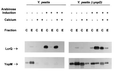
FIG. 5.
Immunoblot analysis of LcrQ and YopM expressed in Y. pestis KIM8-3002 (parent) and Y. pestis KIM8-3002.2 (ΔyopD) with overexpression of LcrQ from plasmid pAW162 in both strains. Bacteria were grown at 37°C in the absence (−) or presence (+) of 2.5 mM Ca2+. Expression of LcrQ from pAW161 was either uninduced (−) or induced (+) by the addition of arabinose to 0.2% (wt/vol). Proteins from equal numbers of cells (0.025 A620 unit · ml) were separated by SDS-PAGE in a 12 (YopM) or 15% (LcrQ) (wt/vol) polyacrylamide gel. Antibodies were used to detect LcrQ and YopM in both the whole-cell (C) (cytoplasmic, periplasmic, and membrane) fraction and the extracellular (E) fraction. The positions of the LCR-related proteins are indicated.

