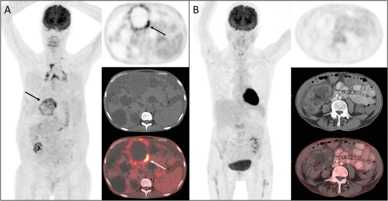
Figure 1:
Representative [18F]-FDG-PET/CT images of CyI according to the visual 4-point scoring system. (A) Maximal intensity projection (MIP) image, transversal PET image of the abdomen, the corresponding transversal CT image and the fusion PET/CT image in a patient with suspected liver CyI, score 4. The arrowhead shows the infected cyst considered for the scoring. (B) MIP image, transversal PET image of the abdomen, the corresponding transversal CT image and the fusion PET/CT image in a patient without suspicion of CyI, score 1.

