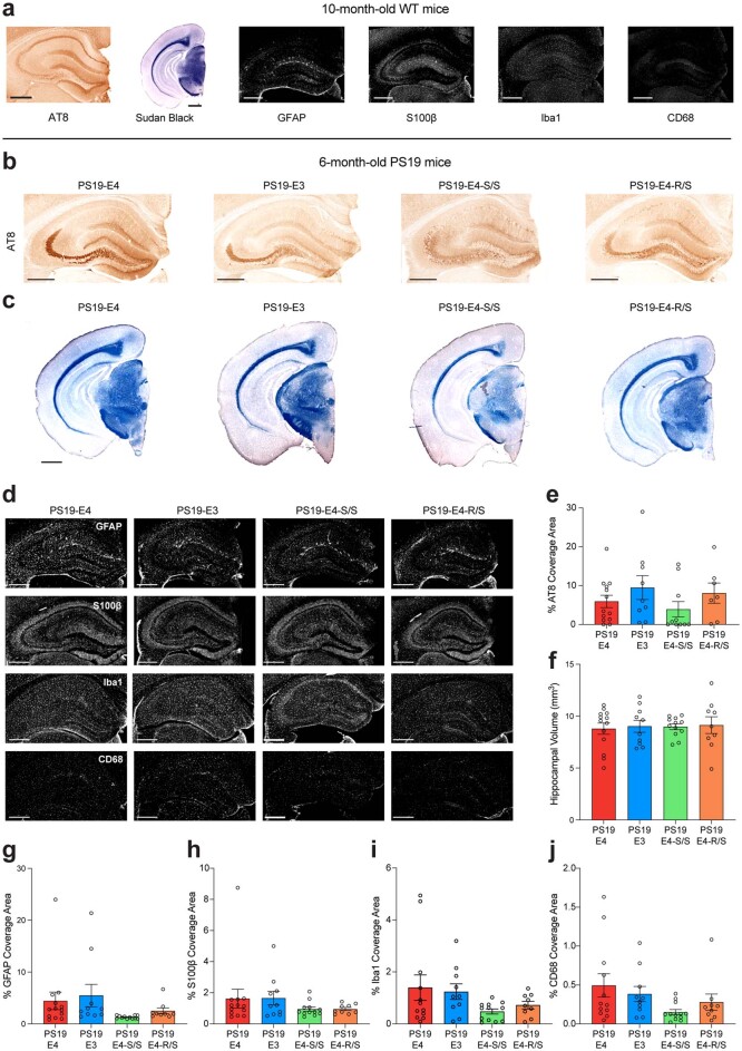
Extended Data Fig. 2. Histopathological analyses of 10-month-old WT and 6-month-old PS19-E3, PS19-E4, PS19-E4-S/S, and PS19-E4-R/S mice.
a, Representative images of 10-month-old WT mouse (n = 11) brain sections stained with AT8 monoclonal antibody to visualize p-Tau (scale bar, 500 µm), Sudan black to enhance hippocampal visualization (scale bar, 1 mm), GFAP and S100β to measure astrocytosis (scale bar, 500 µm), and Iba1 and CD68 to measure microgliosis (scale bar, 500 µm). b,c, Representative images of 6-month-old PS19-E4, PS19-E3, PS19-E4-S/S, and PS19-E4-R/S mouse brain sections stained with AT8 antibody for p-Tau (scale bar, 500 µm) (b) or Sudan black (scale bar, 1 mm) (c). d, Representative images of GFAP and S100β immunostaining for astrocytes and reactive astrocytes, respectively, as well as Iba1 and CD68 immunostaining for microglia and reactive microglia, respective, in the hippocampus of 6-month-old PS19-E4, PS19-E3, PS19-E4-S/S, and PS19-E4-R/S mice. Scale bar, 500 µm. e–j, Quantification of % AT8 coverage area (e), hippocampal volume (f), % GFAP coverage area (g), % S100β coverage area (h), % Iba1 coverage area (i), and % CD68 coverage area (j). In a–j, PS19-E4, n = 12; PS19-E3, n = 11; PS19-E4-S/S, n = 12; PS19-E4-R/S, n = 9; n=mice. Experiments depicted in representative images in a–d were performed using 2 brain sections per mouse, with reproducible data. Throughout, data are expressed as mean ± s.e.m. Differences between groups were determined by ordinary one-way ANOVA followed with Dunnett’s multiple comparison test (e,f) or Welch’s ANOVA followed with Dunnett T3 multiple comparison test (g-j); comparisons of p ≤ 0.05 were labeled on graph.

