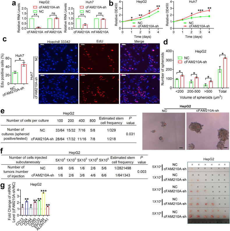
Fig. 6. Knocking down cFAM210A promotes the proliferation, stemness, and tumorigenicity of HCC cells.
a qPCR results showing the expression of cFAM210A and mFAM210A after using cFAM210A-sh lentivirus and its negative control. b CCK-8 assays were performed in cFAM210A-sh HCC cells and their negative controls. c EdU incorporation assays were performed in Huh7 cells (left). Representative images; scale bar, 100 μm (right). d Spheroid formation assays were performed in HepG2 cells (top). Representative images (bottom). e, f In vitro (e) and in vivo (f) limiting dilution assays. The estimated stem cell frequency of each group was calculated by ELDA (http://bioinf.wehi.edu.au/software/elda/); scale bar, 50 μm. g qRT‒PCR showing the expression of seven markers of cancer stem cells in HCC. For (a) and (g), ACTB was used as an endogenous control. For (a–d) and (g), Student’s t test was used. NC negative control, cFAM210A-sh, lentivirus-mediated short hairpin RNA against cFAM210A. ns not significant. *P < 0.05; **P < 0.01; ***P < 0.001.

