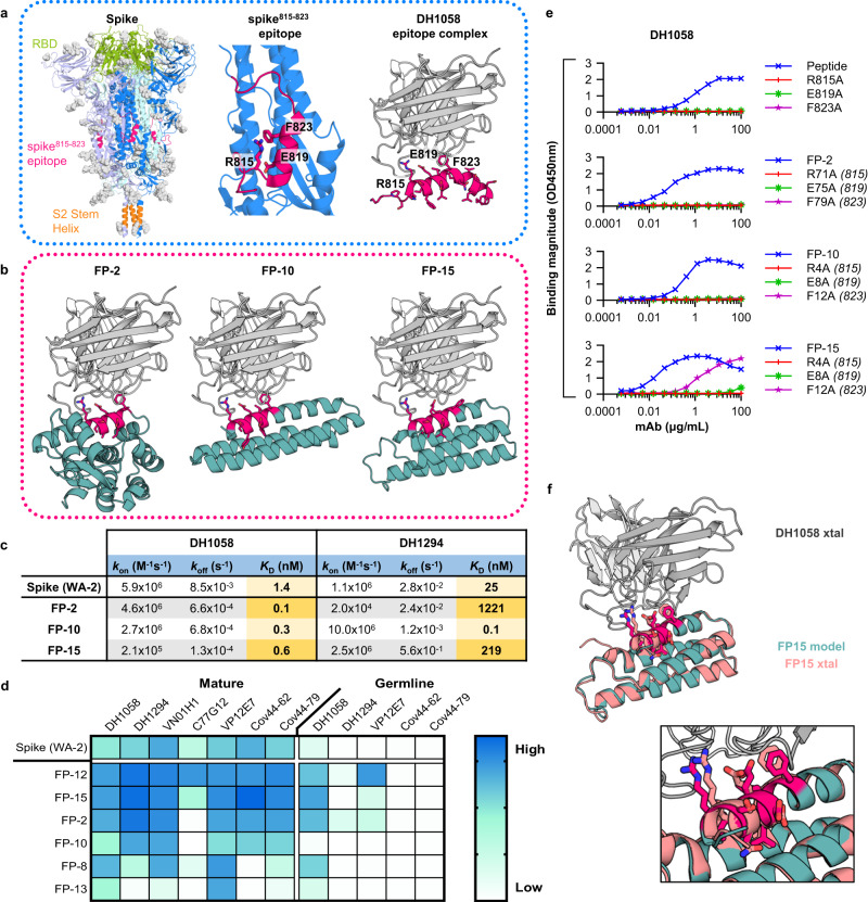
Fig. 1. Design of epitope scaffolds that bind to broadly cross-reactive antibodies against the spike815–823 peptide.
a Left: Structure of the pre-fusion spike trimer (individual monomers: blue, violet and pale green; glycans: gray; PDBid:6xr8), with the Receptor Binding Domain (RBD) (green), spike815–823 peptide (red) and stem helix (orange) domains highlighted. Middle: Zoom of spike monomer with spike815–823 peptide highlighted (red) showing key residues engaged by antibodies. Right: Structure of spike815–823 peptide (red) bound to DH1058 mAb (gray) (PDBid:7tow). b Computational models of DH1058 (gray) bound to ESs FP-2, FP-10 and FP-15 (green) with the grafted epitope shown in red sticks. c Binding affinities of ESs and spike to DH1058 and DH1294 mAbs as determined by SPR. d Binding of spike and ES to diverse spike815–823-targeting mAbs and their inferred precursors. e ELISA binding of DH1058 mAb to synthetic spike815–823 peptides and representative ESs that contain alanine mutations at epitope residues critical for antibody recognition. Numbers in brackets indicate the equivalent position of the mutated epitope residues on spike. f Crystal structure of DH1058 mAb (gray) in complex with FP-15 (salmon) overlaid with the computational model of the ESs (green). Epitope residues are shown in sticks (model: red; crystal structure: salmon). Source data are provided as a Source Data file.

