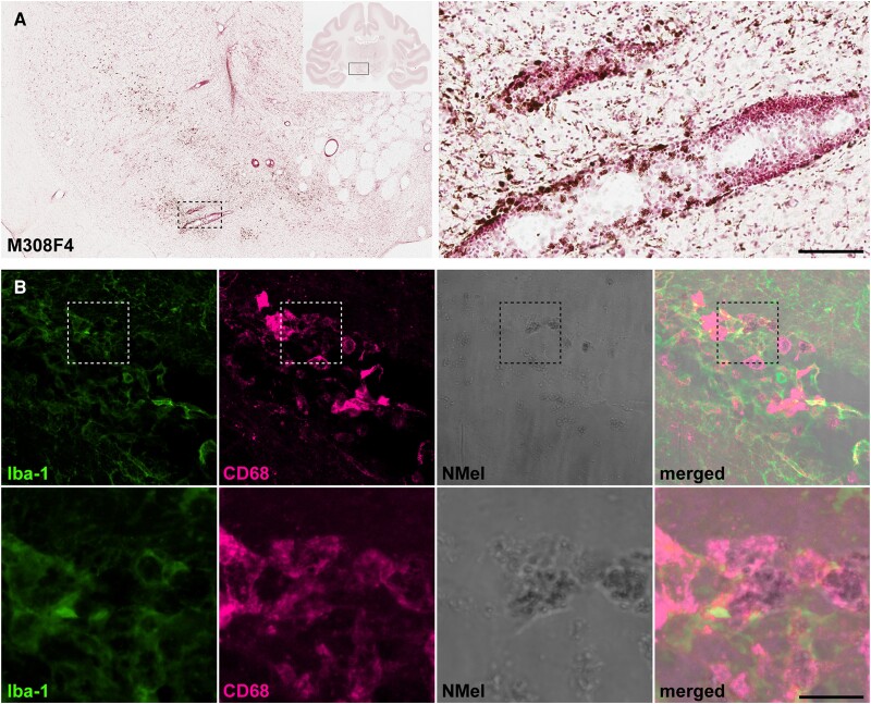
Figure 5.
Extracellular neuromelanin accumulation and pro-inflammatory scenario. (A) Coronal section through the substantia nigra pars compacta (SNpc) in Animal M308F4 counterstained with neutral red and showing the distribution of extracellular neuromelanin resulting from the loss of dopaminergic cells. Inset illustrates the preferential perivascular location of extracellular neuromelanin deposits. Scale bar = 1000 μm (left) and 1000 μm (inset). (B) Confocal images showing the pro-inflammatory scenario made by cells positive for Iba-1 (green channel) and CD68 (purple channel) that are digesting extracellular neuromelanin. Scale bar = 64 μm (A–D) and 18 μm (A’–D’).

