Figure 2.
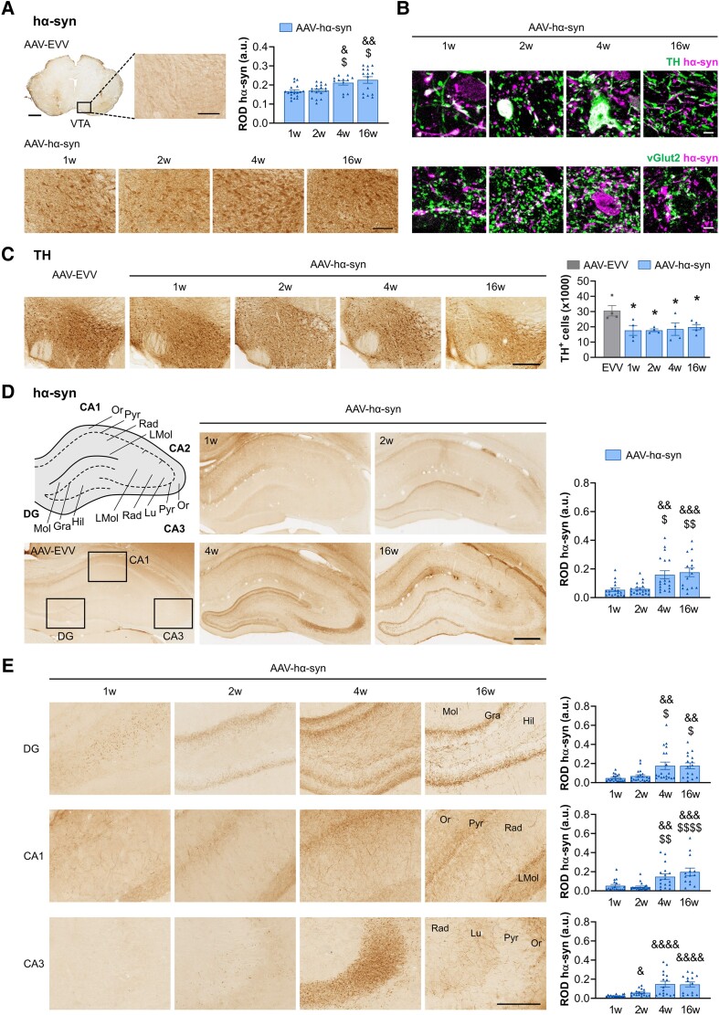
Histological characterization of the VTA and hippocampus. (A) Representative photomicrographs of human α-synuclein (hα-syn) staining in the ventral tegmental area (VTA) in AAV-EVV and AAV-hα-syn rats at 1, 2, 4 and 16 weeks (w) post-inoculation (p.i.). Scale bars: low magnification photomicrographs = 1 mm and high magnification photomicrographs = 100 μm. Relative optical density (ROD) quantification of hα-syn expression. (B) Representative high magnification photomicrographs of hα-syn co-localization with tyrosine hydroxylase (TH) and vGlut2 staining in the VTA. Scale bars = 5 μm. (C) Representative photomicrographs of TH staining in the VTA of AAV-EVV and AAV-hα-syn animals at 1, 2, 4 and 16 weeks p.i. Scale bar = 300 μm. Bilateral quantification of TH+ neurons. (D) Schematic representation of the anatomical regions of the hippocampus. Representative photomicrographs of hα-syn staining in AAV-EVV and AAV-hα-syn animals at 1, 2, 4 and 16 weeks p.i. Scale bar = 500 μm. ROD analysis of hα-syn expression in the whole hippocampus. (E) Representative higher magnification photomicrographs of hα-syn staining in the DG, CA1 and CA3 regions of the hippocampus in the AAV-hα-syn animals at 1, 2, 4 and 16 weeks p.i. Scale bar = 200 μm. ROD analysis of hα-syn expression in DG, CA1 and CA3 regions. The different anatomical regions and layers of the hippocampus are indicated as follows: dentate gyrus (DG)—molecular layer (Mol), granular cell layer (Gra), and hilus (Hil); CA1—stratum oriens (Or), pyramidal cell layer (Pyr), stratum radiatum (Rad) and stratum lacunosum-moleculare (LMol); CA3—Or, Pyr, stratum lucidum (Lu) and LMol. All values are presented as the mean ± SEM and they were analysed using a Kruskal-Wallis test followed by Dunn's post hoc test: &P < 0.05, &&P < 0.01, &&&P < 0.001, &&&&P < 0.0001 versus 1 week p.i.; $P < 0.05, $$P < 0.01, $$$$P < 0.0001 versus 2 weeks p.i.; *P < 0.05 versus AAV-EVV (n = 4 for each group and time point). AAV = adeno-associated viral vector; EEV = empty vector.

