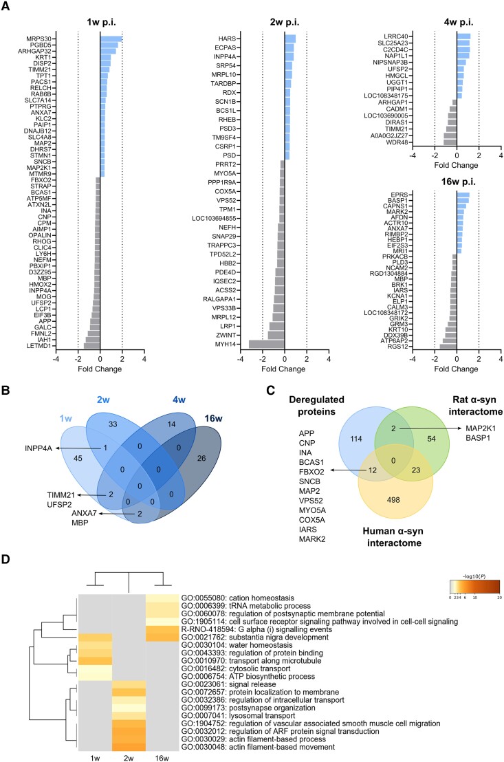
Figure 4.
Deregulated proteins in hippocampal synaptosomes. (A) Differentially expressed proteins (DEPs) at 1, 2, 4 and 16 weeks (w) post-inoculation (p.i.) in the AAV-hα-syn animals compared to the AAV-EVV group. The colours indicate the fold change of the downregulated (grey) and upregulated proteins (blue). (B) Cluster overlap of DEPs at different time points p.i. (C) Cluster overlap between the overall deregulated proteins and the α-syn interactome of both rat and human. (D) Heat map showing the enriched biological pathways and gene ontology clusters at 1, 2 and 16 weeks p.i. No enriched pathways were observed at 4 weeks p.i. The colour scale represents the statistical significance and the grey colour indicates a lack of significance (n = 5 for each group and time point). AAV = adeno-associated viral vector; EEV = empty vector; hα-syn = human α-synuclein.

