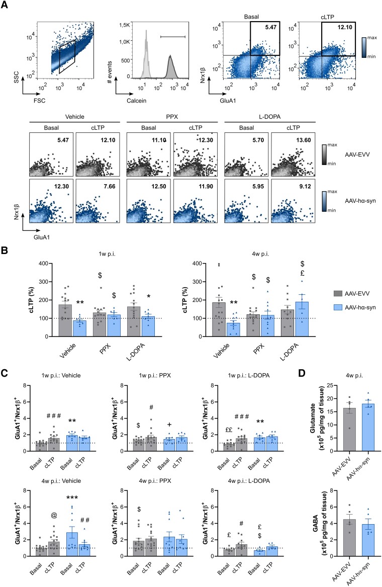
Figure 5.
Flow cytometry evaluation of hippocampal cLTP. (A). Top: Selection of the synaptosomal fraction based on size (FSC) and complexity (SSC). Synaptosomal membrane integrity was evaluated by Calcein AM staining, and GluA1/Nrx1β staining was analysed within Calcein+ events in the basal state and after chemical long-term potentiation (cLTP) stimulation. Bottom: Representative GluA1+/Nrx1β+ double-positive events at the basal state and after cLTP stimulation in the presence of the vehicle (distilled water), pramipexole (PPX) or L-DOPA. (B) The cLTP (%) at hippocampal synaptosomes in the presence of the vehicle (distilled water), PPX or L-DOPA at 1 and 4 weeks (w) post-inoculation (p.i.). (C) GluA1+/Nrx1β+ double-positive values at the basal state and after cLTP stimulation in the presence of the vehicle (distilled water), PPX and L-DOPA at 1 and 4 weeks p.i. GluA1+/Nrx1β+ values were normalized to the AAV-EVV vehicle condition for each time point, and all the values are represented as the mean ± SEM, and assessed using two-way ANOVA followed by a Tukey’s or Bonferroni’s post hoc test: #P < 0.05, ##P < 0.01, ###P < 0.001 versus basal; @P = 0.0616 versus basal; *P < 0.05, **P < 0.01, ***P < 0.001 versus AAV-EVV; $P < 0.05 versus vehicle; +P = 0.0639 versus vehicle; £P < 0.05, ££P < 0.01 versus PPX (n = 8 for each group and time point). (D) Total glutamate and GABA content in the hippocampus at 4 weeks p.i. (n = 5 for each group). The values are represented as the mean ± SEM and assessed with a Mann-Whitney U-test: no significant differences. AAV = adeno-associated viral vector; EEV = empty vector; hα-syn = human α-synuclein.

