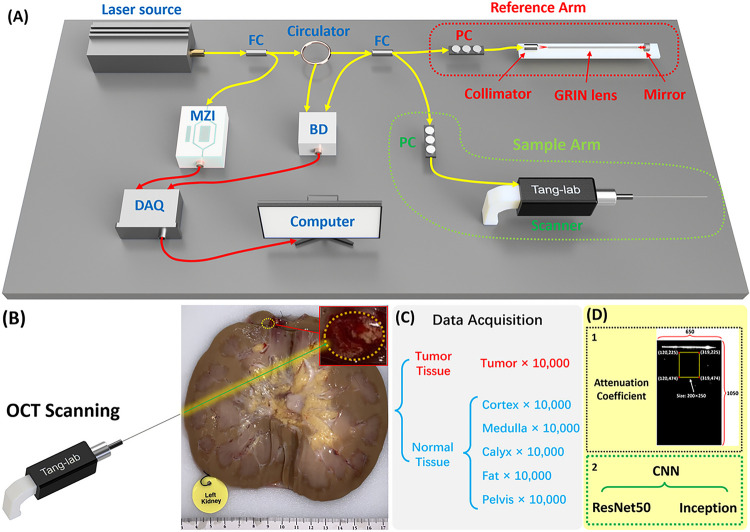
Figure 1.
(A) Schematic of the forward-viewing OCT probe. FC: fiber coupler;PC: polarization controller; MZI: Mach–Zehnder interferometer; BD: balanced detector; DAQ: Data acquisition. (B) Picture of the human kidney with tumor. (C) Data acquisition procedure. (D) Two classification methods used for tissue classification.

