
FIGURE 2.
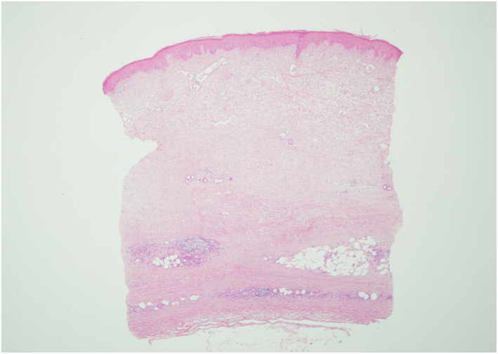
Skin biopsy showing characteristic square shape, abnormal, swollen dermal collagen bundles and a perivascular infiltrate of plasma cells, lymphocytes and histiocytes. The process extends into the subcutaneous fat.
Official websites use .gov
A
.gov website belongs to an official
government organization in the United States.
Secure .gov websites use HTTPS
A lock (
) or https:// means you've safely
connected to the .gov website. Share sensitive
information only on official, secure websites.
Skin biopsy showing characteristic square shape, abnormal, swollen dermal collagen bundles and a perivascular infiltrate of plasma cells, lymphocytes and histiocytes. The process extends into the subcutaneous fat.