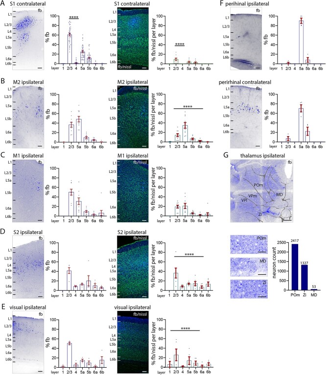
Fig. 2.
Long-range input to S1 L1 from other cortical areas. A to E) Percentage of total fb-labeled neurons (left), and percentage of labeled neurons in relation to nissl in each layer (right). A) S1 contralateral cortex, B) M2 cortex, C) M1 cortex, D) S2 cortex, E) visual (V1, V2L) cortices, F) perirhinal cortices ipsilateral and contralateral. Counts of fb-labeled neurons ipsilateral to application site (left) and contralateral (right). Quantification of the laminar pattern of long-range input to L1 shows the highest percentage of fb-labeled neurons in cortical L2/3 and L5a. G) Fast blue label (total numbers) in POm thalamus, zona incerta, and midline thalamus shows that these 3 subcortical nuclei have projections to L1. Abbreviations in g: MD, midline; POm, posteromedial complex of thalamus; VPl, ventrolateral nucleus of thalamus; VPm, ventromedial nucleus of thalamus; Zi, zona incerta. Each dot in the graphs represents one brain section. Data are from 4 brains. Statistical analysis with 1-way ANOVA, Bonferroni post hoc test, ****P < 0.0001. Analysis details in Supplementary Table 1b. Scale bars in A to F 100 μm, in G 500 μm, in zoom-ins 50 μm.

