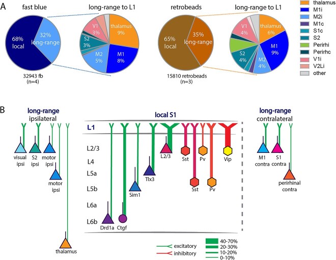
Fig. 7.
Comparison of anatomical input to L1. A) Schematic of input to L1 assessed with fb (left) and retrobeads (right). The bulk of input to L1 was from local neurons and a fraction of the input arising from long-range sources. Thalamus (including POm, zona incerta), ipsilateral and contralateral motor cortices, contralateral S1, ipsilateral S2, bilateral perirhinal ), and ipsilateral visual cortices all provide input to L1. B) Schematic of input to L1. Proportion of local and long-range input and classification of long-range input to L1. Local input arose mainly from L2/3 and L5. Long-range input arose from S2, motor, and visual cortices ipsilateral to the application site and was less pronounced from the contralateral S1 and M1. Abbreviations in a: I, ipsilateral; c, contralateral. Data from 4 mice for fb, 3 mice for retrobeads, analysis details in Supplementary Table 4.

