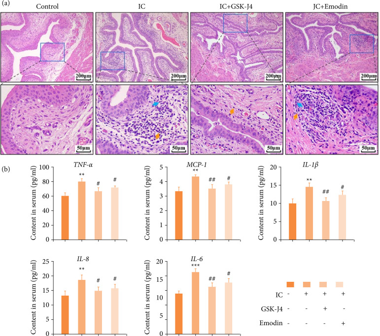
Figure 2. Changes in urinary bladder histology and inflammation after emodin treatment during CYP-induced IC. (a) Histomorphology in four groups of mice urinary bladders was analyzed by hematoxylin and eosin staining. 100× (scale bar = 200 μm) and 400× (scale bar = 50 μm) final magnification. (b) The content of inflammation-related genes (TNF-α, MCP-1, IL-1β, IL-8, and IL-6) in serum, detected by enzyme-linked immunosorbent assay. Data are represented as the mean ± standard deviation.
Source: Elaborated by the authors. IC: interstitial cystitis; TNF: tumor necrosis factor; MCP: monocyte chemoattractant protein; IL: interleukin; blue arrow: neutrophils; yellow arrow: lymphocytes; compared with control, **P < 0.01, ***P < 0.001; compared with IC group, # P < 0.05, ## P < 0.01 represent significant differences between the indicated columns.

