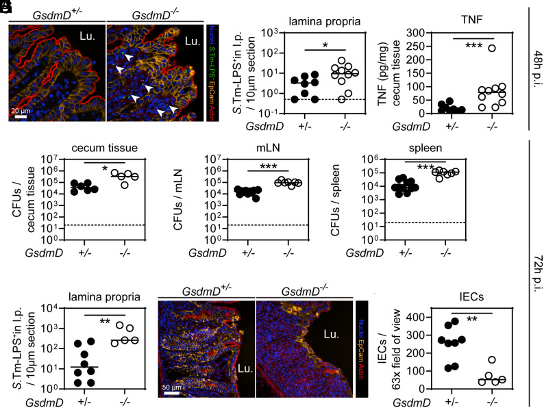
Fig. 2.
GSDMD reduces lamina propria S.Tm loads and protects the gut tissue integrity by 72 h of infection. (A–C) At 48 h p.i., GSDMD-deficient mice exhibit elevated S.Tm pathogen loads in the gut tissue, leading to high levels of TNF compared to heterozygous littermate controls. (A) Representative micrographs of cecum tissue sections, stained for S.Tm-LPS. Arrowheads indicate S.Tm in the lamina propria. Lu. - lumen. (B) Microscopy-based quantification of S.Tm-LPS+ cells in the lamina propria. (C) TNF concentrations in cecum tissue. (D–I) At 72 h p.i., GSDMD deficiency still results in elevated S.Tm pathogen loads locally and systemically, and epithelial tissue integrity becomes compromised. S.Tm CFU pathogen loads in (D) cecum tissue, (E) mesenteric lymph nodes, and (F) spleen. (G) Microscopy-based quantification of S.Tm-LPS+ cells in the lamina propria. (H) Representative micrographs of cecum tissue sections, stained for epithelial marker EpCam. Lu.–lumen. (I) Microscopy-based quantification of IECs per 63× field of view. In B–I, each data point represents one mouse. ≥5 mice per group from ≥2 independent experiments for each comparison. Line at median. The dotted line represents the detection limit. Mann-Whitney U test (*P < 0.05, **P < 0.01, ***P < 0.001).

