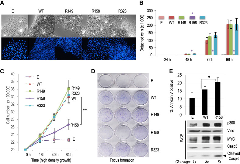
Figure 3.
Cell adhesion, contact inhibition and apoptosis of MYC transformed Rat1a fibroblasts are regulated by the acetylated K158 residue of MYC. (A) Phase contrast (top) and DAPI fluorescence microscopy (bottom) of Rat1a-E and RAT1a-MYC WT and R mutant cell lines. (B) Detachment assay of logarithmically growing cell lines at different times after seeding on regular cell culture plates. The asterisk indicates the lack of significant detachment of Rat1a-MYC/R158 mutant cells (Rat1a-E control cells do not detach). (C) High-density (postconfluency) proliferation assay. Cell lines were confluent at 16 h after plating on regular cell culture plates. (D) Postconfluency growth and formation of dense foci visualized by crystal violet staining. (E) The indicated Rat1a-E or Rat1a-MYC (WT and R158) cell lines were serum-starved, and the fraction of apoptotic cells was determined by annexin V staining or by Western blot analysis of cleaved caspase-3. Cleaved caspase-3 signals were normalized to vinculin and are relative to control (E) cells.

