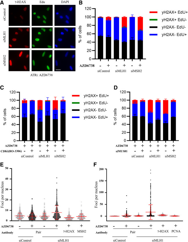
Figure 2.
ATRi preferentially induces DNA damage in MMR-d cells. (A,B) U2OS cells were transfected with control, MHL1, or MSH2 siRNA for 2 d; pulse-labeled with EdU for 20 min; and then treated with 2 µM ATRi (AZD6738) for 4 h. γ-H2AX and biotinylated EdU were analyzed by immunofluorescence. Representative images are shown in A. Fractions of cells positive for γ-H2AX and EdU were quantified as shown in B. Scale bar, 20 µm. (C) U2OS cells transfected with control, MLH1, or MSH2 siRNA were treated with 10 μM CDK1i (RO-3360) for 20 h followed by 2 μM ATRi (AZD6738) for 4 h. Fractions of cells positive for γ-H2AX and EdU were quantified. (D) U2OS cells were transfected with control, MLH1, MSH1, and MUS81 siRNAs as indicated and then treated with 2 μM ATRi (AZD6738) for 4 h. Fractions of cells positive for γ-H2AX and EdU were quantified. Scale bar, 20 µm. (E) U2OS cells were transfected with control or MLH1 siRNA and treated with 2 μM ATRi (AZD6738) for 4 h. Cells were analyzed by PLA using γ-H2AX antibody, MSH2 antibody, or both. The numbers of PLA foci in individual cells were quantified. (Red bars) Mean PLA foci per nucleus in cell populations. (F) U2OS cells were treated as in E and analyzed by PLA using γ-H2AX antibody, PCNA antibody, or both. The numbers of PLAs in individual cell foci were quantified as in E.

