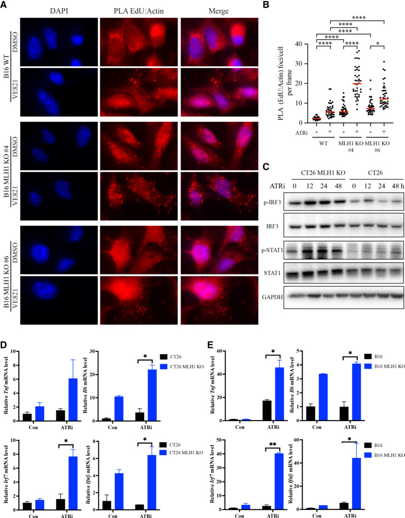
Figure 4.
ATRi increases cytosolic DNA and triggers interferon responses in MMR-d cells. (A,B) B16 or B16 Mlh1 KO cells were labeled with EdU for 2 h and then treated with 10 μM ATRi (VE-821) for 3 h. Cells were analyzed by PLA using antibodies to biotin (EdU) and actin. (A) Images of cells with or without EdU-actin PLA foci. (B) The numbers of PLA foci in multiple image frames were quantified, and the number of PLA foci per cell in each frame was determined (represented as individual dots). (Red bars) Mean number of PLA foci per cell in cell populations. (C) Western blots showing the levels of the indicated proteins and phosphorylated proteins in CT26 and CT26 Mlh1 KO cells after 2 μM ATRi (AZD6738) treatment at the indicated time points. (D,E) The mRNA levels of the indicated interferon-stimulated genes (ISGs) were analyzed by RT-qPCR in CT26 and CT26 Mlh1 KO cells (D) or in B16 and B16 Mlh1 KO cells (E) after mock or 2 μM ATRi (AZD6738) treatment for 2 d.

