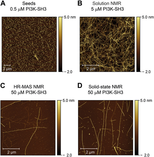
FIGURE 4.

Atomic force microscopy images of PI3K-SH3 fibril seeds and the content of the NMR sample containers at the end of the fibrillation study were monitored by the three different NMR techniques (solution, HR-MAS, solid-state NMR). (A) AFM image of fibril seeds of bovine PI3K-SH3 used in this study. Fibril seeds produced by ultrasonication of PI3K-SH3 fibrils appear as short fibril fragments with approximate main lengths of ∼ 0.3–0.7 µm. Seeds were added at 1% molar monomer equivalent concentration to monomeric PI3K-SH3 for all aggregation experiments. (B–D) AFM micrographs of the PI3K-SH3 fibrils obtained at the end of the solution NMR (B), HR-MAS (C), and solid-state NMR (D) measurements. For AFM measurements, samples were diluted 1:50 for the solution NMR sample and 1:5 for the solid-state NMR and HR-MAS samples performed under MAS conditions. As visible from the AFM images, PI3K-SH3 fibril seeds substantially elongated to fibrils with several µm in length.
