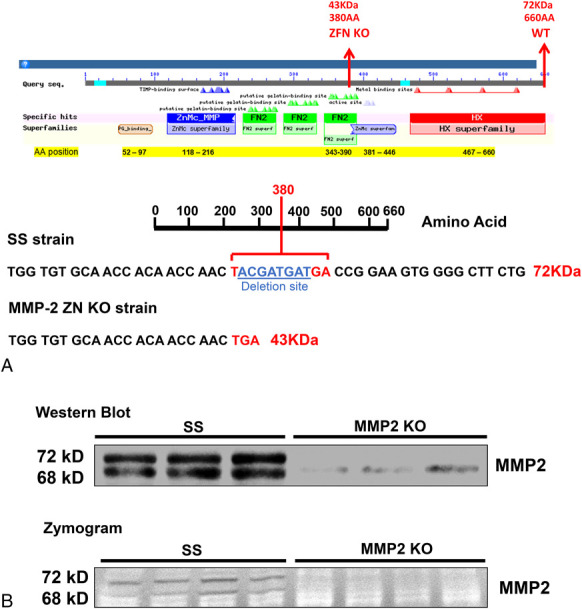
FIGURE 1.

Validation of MMP2 KO using the ZFN technology, Western blot, and zymography. A, Schematic presentation of the MMP2 protein (74 KDa; 660 AA) showing the locations of the different domains and the ZFN-target site that resulted in an 8-bp deletion and the introduction of a premature TGA stop-codon (43 KDa; 380 AA) in MMP2 KO rats. B, Representative images of Western blot and zymography confirming the loss of the expression of MMP2 protein and activity in the kidney of MMP2 KO rats. AA, amino acids; FN2, fibronectin type II repeats (green); HX, hemopexin-like domain (red); ZnMc, zinc binding site (blue).
