
FIGURE 6.
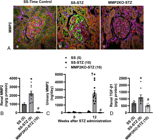
Effect of MMP2KO on the renal expression and urinary excretion of MMP2 in STZ-treated diabetic SS rats. A, Representative images of the renal cortex stained with MMP2 antibody (green) and counterstained with 0.001% Evans blue (red) and DAPI (blue) in age-matched control SS rats (SS), STZ-treated SS (SS-STZ), and MMP2 KO (MMP2 KO-STZ) rats. Magnification, ×200. MMP2 was avidly stained in the glomeruli, proximal tubules, and interstitial space of (B) SS-STZ rats compared with (A) SS control rats. There was no staining of MMP2 in the kidney of (C) MMP2 KO-STZ rats. B, Expression of MMP2 protein in the renal cortex of SS, SS-STZ, and MMP2 KO-STZ rats measured with ELISA 12 weeks after induction of diabetes. C, Urinary excretion of MMP2 was measured in SS, SS-STZ, and MMP2 KO-STZ rats before and 12 weeks after induction of diabetes with STZ. D, Protein levels of TGF-β1 in the renal cortex of SS, SS-STZ, and MMP2 KO-STZ rats were measured with ELISA 12 weeks after induction of diabetes. Mean values ± SEM are presented. Numbers in parentheses indicate the number of rats studied per group. * indicates P < 0.05 versus the corresponding value measured in SS control rats. # indicates P < 0.05 versus the corresponding value measured in SS-STZ rats. † indicates P < 0.05 versus the corresponding value measured within the same strain at week 0.