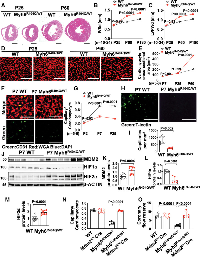
Figure 6.
MDM2 regulates capillary formation in Myh6R404Q/WT mice before the development of ventricular hypertrophy. A, Representative hematoxylin-eosin–stained heart cross-sections of postnatal days 25 (P25) and 60 (P60) WT and Myh6R404Q/WT mice. Scale bars=1 mm. Echocardiography assessment of (B) interventricular septal thickness at end diastole (IVSd) and (C) left ventricular posterior wall thickness at end diastole (LVPWd) in WT (n=10–11) and Myh6R404Q/WT (n=19–24) mice at P25, P60, or postnatal day 180 (P180). D, Representative images of WGA (red)–stained left ventricular tissue from WT and Myh6R404Q/WT mice at P25 or P60. Scale bar=75 μm. E, Cardiomyocyte cross-sectional areas from WGA-stained left ventricular tissue from WT (n=5–6) and Myh6R404Q/WT (n=5–6) mice at postnatal day 7 (P7), P25, or P60. Minimum 50 cardiomyocytes per sample. F, Representative immunohistochemistry images for CD31 (green) costained with WGA (red) in left ventricular tissue from P7 WT and Myh6R404Q/WT mice. Nuclei are blue (DAPI). Scale bars=50 μm. G, Capillary-to-cardiomyocyte ratios in left ventricular tissue from WT (n=5–6) and Myh6R404Q/WT (n=5–6) mice at postnatal day 2, P7, or P25. Minimum 200 cardiomyocytes per sample. H, Representative fluorescence images for the intravascularly injected endothelial cell stain T-lectin (green) in left ventricular tissue from P7 WT and Myh6R404Q/WT mice. Scale bars=80 μm. I, Capillaries per mm2 in left ventricular tissue from P7 WT (n=6) and Myh6R404Q/WT (n=6) mice; 3 cross-sectional images per sample were analyzed. J through M, Immunoblots and quantification for MDM2, HIF1α, and HIF2α in left ventricular tissue lysates from P7 WT (n=6–9) and Myh6R404Q/WT (n=6–9) mice normalized to β-actin and relative to WT. N, Capillary-to-cardiomyocyte ratios were calculated from left ventricular tissue in P7 WT (n=6), Mdm2fl/+/Myh6:Cre (n=6), Myh6R404Q/WT (n=6), and Myh6R404Q/WTMdm2fl/+/Myh6:Cre (n=6) mice. Minimum 200 cardiomyocytes per sample. O, Coronary flow reserve in P25 WT (n=5), Mdm2fl/+/Myh6:Cre (n=5), Myh6R404Q/WT (n=6), and Myh6R404Q/WTMdm2fl/+/Myh6:Cre (n=5) mice. All results are shown as mean±SEM. Student or Welch t test used for I, K, L, and M; 2-way ANOVA with Tukey multiple comparison test used for B, C, E, G, N, and O. CD31 indicates cluster of differentiation 31; DAPI, 4′,6-diamidino-2-phenylindole; HIF1α, hypoxia-inducible factor 1 alpha; HIF2α, hypoxia-inducible factor 2 alpha; MDM2, murine double minute 2; Mdm2fl/+Cre, Mdm2 heterozygous floxed and Myh6:Cre; Mybpc3-/-, cardiac myosin binding protein 3 homozygous deletion; Myh6:Cre, myosin heavy chain 6:Cre recombinase; Myh6R404Q/WT, myosin heavy chain 6 arginine to glutamine substitution at amino acid 404 heterozygous; T-lectin, tomato lectin; WGA, wheat germ agglutinin; and WT, wild-type.

