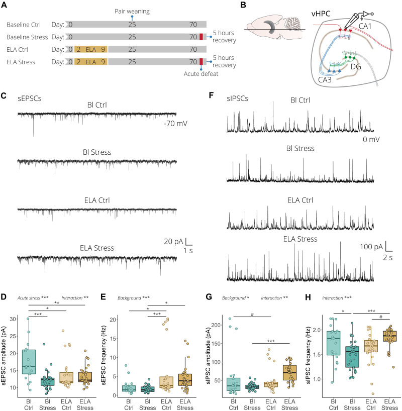
Fig. 5. ELA alters excitation and inhibition transmission in response to acute stress in the vHPC.
(A) Experimental timeline for the electrophysiological recording of the baseline control, baseline stress, ELA control, and ELA stress conditions. (B) Illustration of an ex vivo acute ventral hippocampal brain slice depicting whole-cell patch-clamp recording obtained from CA1 pyramidal neurons. (C) Representative spontaneous excitatory postsynaptic current (sEPSC) recordings from CA1 pyramidal neurons. (D) Quantification of sEPSC amplitude. Acute stress reduces sEPSC amplitude, while ELA-exposed animals have chronically reduced sEPSC amplitude with no further effect of acute stress (left). (E) In wild-type mice, acute stress has no effect on sEPSC frequency, while in ELA mice it increases sEPSC frequency (right). (F) Representative spontaneous inhibitory postsynaptic current (sIPSC) recordings from CA1 pyramidal neurons. (G) Quantification of sIPSC amplitude showed that acute stress had no effect in wild-type animals, while acute stress significantly increased sIPSC amplitude in mice with ELA background (left). (H) Acute stress significantly reduced sIPSC frequency in wild-type mice, while the frequency was increased in response to acute stress in ELA mice (right). sEPSCs and sIPSCs were recorded at –70 and 0 mV, respectively, from n = 17 to 30 cells/four to six mice per group. Box plots represent the 25%, median, and 75% quartile; whiskers span 1.5 × IQR. Two-way ANOVA with Tukey post hoc test. #P < 0.1, *P < 0.05, **P < 0.01, ***P < 0.001.

