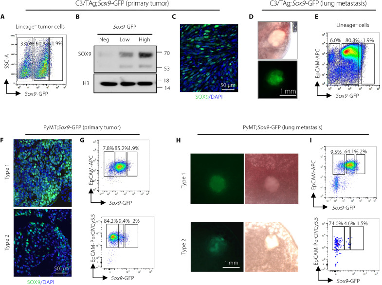
Fig. 1. Sox9-GFP reporter identifies cancer cells expressing distinct levels of SOX9 in spontaneous primary tumors and metastases.
(A) Flow cytometry showing the expression of Sox9-GFP in C3/TAg;Sox9-GFP tumors (n = 22). CD31, CD45, and Ter-119 were used as lineage markers. (B) SOX9 Western blot of cells sorted from C3/TAg;Sox9-GFP tumors as in (A). One representative experiment of three repeats was shown (n = 3). (C) Representative immunofluorescence of SOX9 in C3/TAg;Sox9-GFP primary tumors (n = 3). (D) Representative images of lung metastases in C3/TAg;Sox9-GFP mice (n = 4). (E) Flow cytometry showing the expression of Sox9-GFP in lung metastases in C3/TAg;Sox9-GFP mice (n = 4). (F) Representative immunofluorescence of SOX9 in PyMT;Sox9-GFP tumors (type 1: n = 6; type 2: n = 2). (G) Flow cytometry measuring Sox9-GFP in PyMT;Sox9-GFP tumors (type 1: n = 6; type 2: n = 2). (H) Representative images of lung metastases in PyMT;Sox9-GFP mice (type 1: n = 4; type 2: n = 2). (I) Flow cytometry measuring Sox9-GFP in lung metastases from PyMT;Sox9-GFP mice (type 1: n = 4; type 2: n = 2). DAPI, 4′,6-diamidino-2-phenylindole; PerCP, peridinin-chlorophyll-protein; APC, allophycocyanin; SSC, side scatter; EpCAM, epithelial cell adhesion molecule.

