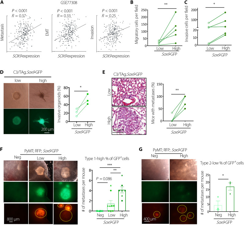
Fig. 3. SOX9high tumor cells have increased metastasis-initiating capacity.
(A) Correlation between SOX9 levels and functional states in breast tumors at the single-cell level as determined by CancerSEA. EMT, Epithelial-Mesenchymal Transition. (B) Transwell migration assay of C3/TAg primary tumor cells (n = 5 tumors). (C) Transwell invasion assay of C3/TAg primary tumor cells (n = 5 tumors). (D) Representative images and frequencies of organoids with the invasive morphology in collagen gel (n = 3 tumors). (E) Lung metastasis-initiating ability of Sox9low and Sox9high C3/TAg primary tumor cells by tail vein injection. Each line indicates one experiment with a distinct tumor (n = 4). (F) Lung metastasis-initiating ability of Sox9neg, Sox9low, and Sox9high PyMT tumor cells by tail vein injection. Metastatic nodules were determined after 14 weeks (triangles and circles representing two different donors). (G) Lung metastasis-initiating ability of Sox9neg, Sox9low, and Sox9high PyMT tumor cells by tail vein injection. Metastatic nodules were determined 14 weeks after injection (n = 3). All data are represented as means ± SEM. P values were determined by two-tailed t test [(B), (C), (D), (E), and (G)] or one-way ANOVA with Tukey’s test (F). ***P < 0.001, **P < 0.01, and *P < 0.05.

