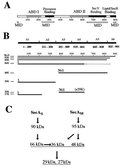
FIG. 7.
Summary of identification and characterization of SecA fragments. (A) Schematic presentation of proposed SecA domains. The two ABDs of SecA (ABD I and ABD II) (25) are shown as shaded rectangles, and the domains interacting with precursor (23, 37) or with lipid and SecB (2) are shown as solid rectangles, on the SecA sequence. The known and proposed MID are shown by open bars below the SecA sequence. (B) Schematic presentation of the identification of SecA fragments. The identified SecA fragments and the six SecA fragments fused to maltose-binding protein to produce the region-specific antibodies are aligned with the SecA sequence illustrated in panel A. Starting amino acid numbers are given for the major fragments of the integral SecA. The open box represents the potential cleavage fragment corresponding to the reported 30-kDa fragment starting at Leu610 (27) but not identified here. (C) Proposed proteolysis pathway for SecAS and SecAM. SecAS, membrane-integral SecA with a conformation similar to that of soluble SecA; SecAM, membrane-integral SecA with a membrane-induced conformation.

