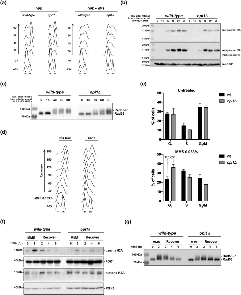
Fig. 2.
Cells lacking Opi1 show a delayed G1-to-S-phase transition and activation of the DDR upon genotoxic stress induced by MMS. a) Flow cytometry analysis depicting S-phase progression in wild-type and opi1Δ strains. b) Western blot analysis of gamma-H2A (histone H2A phosphorylation) in the indicated strains. c) Western blot analysis showing the MMS-induced Rad53 phospho-shift in the indicated strains. d) Flow cytometry analysis illustrating the resumption of S-phase progression in wild-type and opi1Δ strains after MMS treatment (0.033% for 2 h). e) Quantification of the percentage of cells in the G1, S, and G2/M phases in untreated cells and after MMS treatment (0.033% for 2 h). The results shown are representative of 3 independent experiments, and error bars indicate the standard deviation. Two-way ANOVA with Tukey's post hoc testwas used for significance analysis between samples. f) Western blot analysis of gamma-H2A and total histone H2A in the indicated strains. g) Western blot analysis showing the MMS-induced Rad53 phospho-shift in the indicated strains. Note: For a) to c), cells were arrested in G1 after 2-h treatment with α-factor and then released in fresh YPD medium ± 0.033% MMS. All strains are bar1Δ. For d) to g), cells were treated with 0.033% MMS for 2 h and then released in fresh YPD medium. Western blots were probed with anti-gamma-H2A, anti-histone H2A, anti-Rad53, and anti-PGK1 (loading control) antibodies as described in Materials and methods.

