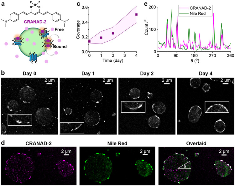
Figure 2.
SMLM with CRANAD-2 indicates the gradual formation of amyloid fibril nanoaggregates at the FUS condensate surface. a. CRANAD-2 chemical structure and mode of action: CRANAD-2 is nonfluorescent in the aqueous phase but turns on fluorescence upon binding to amyloid fibrils in the condensates. b. Representative SMLM images of CRANAD-2, for FUS condensates aged for different days. Insets: Zoom-ins highlighting the surface nanodomains. c. Time-dependent surface coverage of CRANAD-2 SMLM signal at FUS condensates over aging. Data points and shades present the averages and standard deviations of individual condensates (~10 for each data point), respectively. d. Two-color SMLM for a FUS LLPS sample loaded with both CRANAD-2 and Nile Red, shown as separate (CRANAD-2: magenta, Nile Red: green) and overlaid (colocalization: white) images. e. Line profiles of single-molecule localizations in the two dye channels along the circumference of a condensate droplet, presented as counts per degree as a function of the azimuth angle θ marked in (d).

