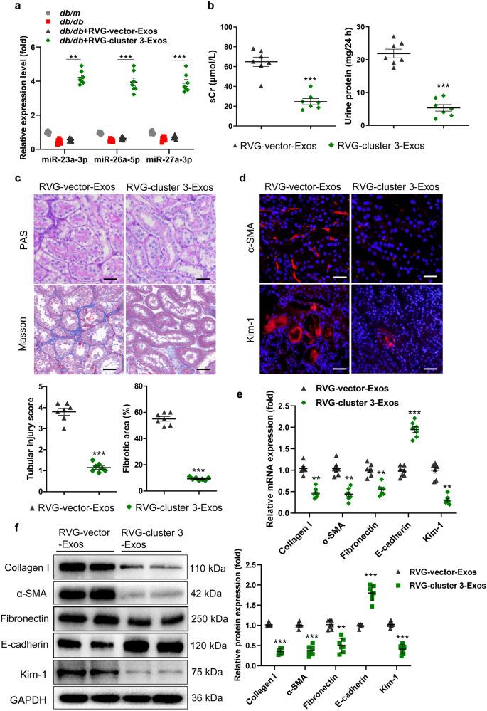
Fig. 3. RVG-miR-23a/27a/26a-Exos attenuate TIF in the mice with DN.
At 12 weeks of age, db/db mice were intravenously administered Exos (100 µg) once a week for 8 weeks until they were sacrificed at 20 weeks of age. a The expression of miR-23a-3p, miR-26a-5p and miR-27a-3p in the kidney were assayed by qRT‒PCR. The results are normalized to U6. n = 7 per group. b sCr and 24 h urinary protein levels. n = 7 per group. c Representative images of PAS and Masson staining of the renal cortex. Scale bars, 20 μm. d Representative images of α-SMA and Kim-1 immunostaining. Scale bars, 20 μm (α-SMA). Scale bars, 50 μm (Kim-1). e, f qRT‒PCR and Western blot analyses of α-SMA, collagen I, Fibronectin, E-cadherin, and Kim-1 expression. The relative levels were normalized to GAPDH. n = 7 per group. **P < 0.01, ***P < 0.001. α-SMA, α-smooth muscle actin; Kim-1, kidney injury molecule-1; TIF, tubulointerstitial fibrosis.

