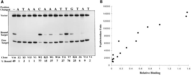
FIG. 2.
(A) Binding of NAC-binding site mutants from hutUp analyzed by gel mobility shift assay. The mutant NAC-binding sites were labeled with 32P and incubated with NAC protein (0.28 μM). Bound and unbound fragments were separated by polyacrylamide gel electrophoresis. The radioactivity in each band was measured as described in Materials and Methods. (B) Correlation between protein binding and transcriptional activation for hutUp NAC-binding site mutants. The graph plots the activated levels of β-galactosidase (grown under nitrogen limitation) from Fig. 1B, and the binding percentages (normalized to clone V16 which was 49%) for the analyzed NAC binding-site mutants from panel A.

