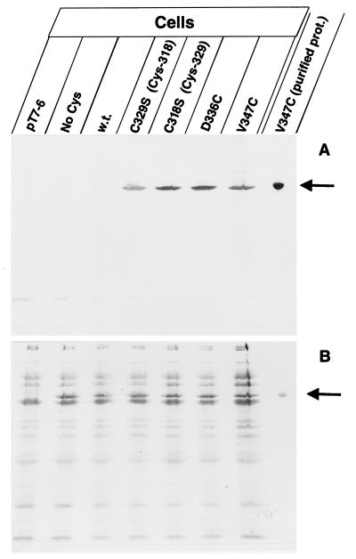
FIG. 2.
Specific labeling of the FhuA single-cysteine mutants with B-M in live cells (A). Cells of E. coli UL3 fhuA were transformed with one of the following plasmids: pT7-6 (vector without fhuA), pfhuA9 [FhuA(C318S C329S C692S C698S); No Cys], pfhuA8 [FhuA(wild type); w.t.], pfhuA5 [FhuA(C329S) Cys-318], pfhuA4 [FhuA(C318S) Cys-329], pfhuA6 [FhuA(D336C)], and pfhuA7 [FhuA(V347C)], as indicated. Cells were incubated for 30 min with 0.5 mM B-M at 30°C, and the proteins of whole cells were separated by SDS-PAGE. In addition, isolated FhuA(V347C) protein was heated for 3 min in 4% SDS and subsequently labeled with B-M (purified prot.). The proteins were separated by SDS-PAGE, blotted onto a nitrocellulose membrane, and stained with Ponceau-S-Red (B) and, after destaining and incubation with streptavidin-β-galactosidase, stained with X-Gal (A). The FhuA band is indicated by an arrow.

