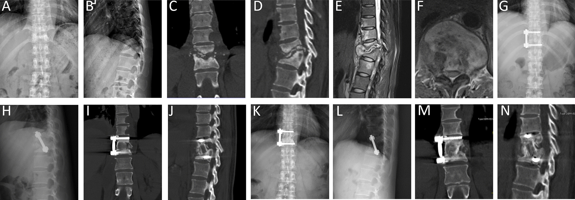
Fig. 2.

A 45-year-old woman with T11–12 tuberculosis. A, B Preoperative X-ray before surgery demonstrated vertebral bone destruction and kyphosis at T11–12. C–F Preoperative CT and MRI showed T11–12 vertebral bone destruction with paravertebral and intraspinal abscess. G–J Postoperative X-ray and CT of the patient underwent anterolateral debridement and instrumentation at T11–12 with autologous structural iliac bone graft. K–N X-ray and CT at 25 months postoperative showed good bone fusion and spinal alignment without signs of tuberculosis recurrence and hardware failure
