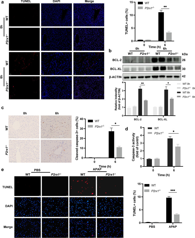
Fig. 2.
P2rx1 depletion eliminates APAP-induced cell death. (a) Representative images of TUNEL staining (original magnification × 200) and the statistical quantification of TUNEL-positive cells in liver sections of WT and P2rx1−/− mice with or without APAP treatment (n = 4–6 per group); (b) Western blot and quantification analysis for the expression of hepatic BCL-2 and BCL-XL in WT and P2rx1−/− mice with or without APAP treatment (n = 3–4); (c) Representative IHC staining (original magnification × 200) and the statistical quantification of hepatic cleaved-caspase-3-positive cells in liver sections of WT and P2rx1−/− mice with or without APAP treatment (n = 4–6 per group); (d) Caspase-3 activity in the livers with or without APAP treatment (n = 4–6 per group); (e) Representative images of TUNEL staining (original magnification × 200, scale bar = 353 μm) and the quantification of TUNEL-positive primary hepatocytes with or without 5 mM APAP treatment in vitro (n = 3–6 per group). Data are shown as the means ± SEM, *p < 0.05, **p < 0.01, ***p < 0.001

